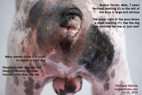
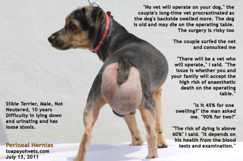
The Boston Terrier was operated one year ago. I review this case which was operated successfully (no anaesthetic deaths or complaint of recurrence) before I operate on the “mother of all perineal hernias” of the Silkie Terrier (described below) who is 10 years old and in a more risky anaesthetic position.
The pictures and more details of this record are at:
http://www.bekindtopets.com/animals/20100625constipation_perineal_hernia_old_Boston_Terrier_male_singapore_ToaPayohVets.htm
CASE 1
This Boston Terrier case was written: 25 June, 2010
Boston Terrier, Male, Not neutered, 7 years, 12.4kg
Big swellings besides the anus, the left swelling being bigger.
Difficulty in pooping for past 3 weeks.
A knowledgeable young man who has his own views of dog care.
He presented a Boston Terrier with constipation for the past 3 weeks. He went to Vet 1 who referred him to another vet after taking a blood test as she did not want to perform the surgery. She had given him a laxative for the dog and the dog had passed loose stools instead of hard ones.
“Why don’t you see the referred vet?” I asked.
“The Surgery has a bad reputation,” the young man had googled the name of the practice he was referred to by Vet 1. “There is a very bad complaint about the vets from one dog owner. So I better not go there.”
“All veterinary practices will have one or two nasty complaints about service,” I educated this young man. “The busier the practice, the higher the chances of getting complaints. This is because the vet has no time to handle each case as thoroughly as he or she would love to. This applies to over-worked doctors in human medicine too especially in the emergency wards.”
VACCINATION HISTORY MUST BE ASKED
“Has your dog been vaccinated?” I asked.
“No vaccination for the past few years. Do you have parvovirus in your Surgery?” the young man asked me.
“Fortunately for your dog, my practice does not have parvo-viral cases for many months as I seldom provide service to the dog breeders nowadays. You have taken a big risk exposing your dog the risk of parvoviral and canine distemper infections.”
PRE-OP BLOOD TEST IMPORTANT FOR OLD DOGS BEFORE SURGERY
I checked Vet 1′s blood test results. It is wise not to trust the blood results of other practices based on one of my experiences (see one case I had written). However, he had paid $ 130 for the test and I would not insist as that would increase his vet bills. Overall, the dog was examined and was healthy. So I took the chance.
ANAESTHESIA
IV saline given. Then I gave Atropine 0.4 ml IV followed by Domitor 0.2 ml IV
Waited 10 minutes. Isoflurane gas mask. Dog struggled. So, I gave Zoletil 100 @ 0.1 ml IV. This sedated the dog who was masked and given isoflurane gas at 5%. The dog slept and was intubated. Isoflurane at 1-2% maintenance was done by my experienced assistant, Mr Saw. I asked him to increase the dose when the pelvic fat kept coming out from the hernia after pushing the fat into the abdominal cavity. The dog recovered smoothly.
SURGERY
I gave him antibiotics to take and schedule perineal hernia surgery 2 days later. The surgery took nearly an hour as the hernia was large. The hernia bulge with pelvic fat is large, around 4 cm x 6 cm. An electro-incision made a big cut to the left of the anus.
It was difficult to identify the medial coccygeal and levator ani muscles in this case as there is a lot of inflammation. The internal pudendal artery and vein and the pudendal nerve on the dorsal surface of the internal obturator muscle looked compressed as I showed to my assistant Mr Saw who nodded his head. Judging from his eyes, he did not believe they were what I said.
Is there a right perineal hernia too?
Electro-incision. Big amount of pelvic fat. A retractor enabled me to have a good field of view to stitch up the defect
See the big hole through which part of the colon and pelvic fat herniated through causing a big backside swelling
Left perineal hernia repaired. Neutering in 3-4 weeks if the owner wants to do it. The right perineal hernia may need to be repaired later.
The internal obturator muscle is on the ventral aspect of the pelvic diaphragm. This was a big fatty mass horizontally covering the muscle, unlike the no-fat muscles illustrated in Small Animal Surgery, T.W. Fossum 1997, pg 354.
I used a retractor to spread open up the operating area and to see the pudendal vessels and nerve just above the obturator muscles in this case. Do not stitch these vessels or nerves.
POST-OP
Dog woke up fast. Given tolfedine painkillers.
TWO HERNIAS TO BE OPERATED AT ONE GO
I doubt that it is possible to do two hernias at one go as the muscle stitching on one side (i.e. left hernia in this case) pulled the left anal area tightly to cover the herniated hole. Therefore doing two hernia repair at the same time just is not in the interest of the dog as he will feel very uncomfortable and painful.
LOOSE STOOLS leaking out from the anus. This must be plugged. The dog had been given an oil laxative by Vet 1 for 3 days and the loose stools start to come out despite atropine injection.
CONCLUSION
The dog was OK and was warded for at least 4 days as the owner did not have a crate to prevent the dog running loose. I checked the dog every day to ensure that he had proper nursing care and pain-killers. The boy’s parents came to visit the dog yesterday. The dog should be back home after 7 days. He had managed to rub his backside onto the floor of the crate despite tolfedine 60 mg at half a tablet per day for 3 days. I decided to give him 1/4 dose of a 30mg phenobarb and then Rimadryl for another 3 days to prevent pain and inflammation.
P.S
1. Yearly vaccination is important. Fortunately this dog did not get infected with parvoviral disease in the practice of Vet 1 which is a very busy practice and in my surgery. Otherwise, I end up with a dog passing blood in the stools and dying later. At the time of writing this report, it is still early at Day 5 after visiting Vet 1. Parvoviral signs come in around 10-14 days after infection.
2. “Neutering the dog when he was younger would have decreased the chances of him getting perineal hernia,” I said. “Perineal hernia is more common in non-neutered dogs.” The young man said: “It is cruel and that is why I don’t do it.” He has been advised to neuter the dog around 2-4 weeks later. As for the right perineal hernia, it is a smaller one. Wait and see. If the dog is neutered and there is
no more swelling in the backside, then there is no need to do a right perineal hernia repair.
3. High anaesthetic risks. I don’t enjoy doing high anaesthetic risk surgeries as they are very stressful for me. If the dog survives, everybody is pleased. There will be deaths and the owners may be very emotional and angry. Some may post a nasty complaint in the internet. To minimise risks of deaths of old dogs on the operating table, I don’t force myself to perform hernia repair and neutering at the same time. The owner has to appreciate that I don’t take risks unnecessary.
UPDATE AS AT JULY 11, 2011
No news from the owner since the surgery in Jun 2010. I presume all are OK as now news is good news.
CASE 2
ANOTHER MORE CHALLENGING CASE ONE YEAR LATER
THE MALE DOG HAD A VERY LARGE BACKSIDE SWELLING – PART 1
Dr Sing Kong Yuen, BVMS (Glasgow), MRCVS
Case: Updated: 14 July, 2011
This Silkie Terrier case was written: 24 June 2011
Silkie Terrier, Male, Not neutered, 1- years, 6.5kg
Big swellings besides the anus, the left swelling being bigger.
Difficulty in pooping for past 3 weeks.
I have not seen perineal hernias since I operated on the above-mentioned Boston Terrier one year ago. Surprisingly, on Sunday, July 10, 2011, my assistant Mr Min said that a couple insisted on seeing me. Normally, all cases go to Dr Vanessa Lin but I was around at the reception to get a pulse of the grass-roots from 9 am to 6 pm and to ensure that the waiting times are kept to the minimum. Only at the receptionist’s counter can I know what is the situation of the waiting time like, rather than depend on the receptionist to enlighten me.
The couple said to me: “My vet said that no vets in Singapore would operate on my 10-year-old Silkie Terrier. He had said that the dog is old and to leave the swelling alone. But it kept growing bigger!”
He was not neutered and had a backside lump 3/4 the size of the biggest mango you can see in Singapore. The dog had difficulty in pooping and the older parents were concerned about his quality of life. The dog could eat, drink, poop and pee without difficulty and was active.
“There will be vets in Singapore that will operate on this dog,” I said when the couple brought the dog in later for examination. “The main issue is that the operation will take a long time and the dog may just die on the operating table. No vets want to do a dog that dies on the operating table.”
I had asked them to illustrate as they did not bring the dog down at first. A male dog, not neutered, big swelling from below the anus (in this case) instead of to the sides of the anus as in unilateral perineal hernias. The couple had actually diagnosed perineal hernias via the internet education and so they knew what was wrong with their dog. The problem was that their vet did not want to operate and put his reputation on the line when the dog dies on the operating table. What should I do?
This was the “mother” of all perineal hernias. Both left and right perineal hernias have “amalgamated” to form a large 3/4-Taiwanese mango-sized lump to the right, left and below the anus. Taiwanese mangoes are gigantic but are not sweet and they measure around 25 cm x 10 cm x 7 cm. So, you can imagine that the swelling was really gigantic.
The surgery will take over 1.5 hours and the old dog’s heart may just stop beating. The dog needs to be operated as he has had been licking his skin thin. Continuous licking to relieve his pain and irritation as the intestines and omental fat prolapse through the pelvic muscular defect from both the left and right side. It was hard for him just to sit down too.
http://www.kongyuensing.com/pic/20110713perineal-hernias-bilateral-silkie-terrier-male-10-years-toapayohvets-singapore.jpg
“Do your parents know that they may not see the dog alive once he gets operated?” I asked. The aged parents are the care-givers. The couple said: “My parents say it is better to take the risk rather than let the dog suffer with such a big dangling mass. The groomer had nicked the lump earlier and discovered this hernia. Otherwise, we would not know it exists!”
The dog was in good body condition. I checked his heart. His heart was surprisingly normal. He was alive and active now. As if he has not a worry in this world while his caregivers bear the burden and surgical risks on their shoulders. Should I pass the buck? And to whom? To my two associate vets? This is the type of challenging cases that I prefer not to take on and it will be most unkind to pass the buck to my two associate vets as there is the possibility of post-surgical complications like infections, bleeding and nerve damages in addition to death on the operating table. So, I did not refer the case to them. It is a moderately difficult surgery but it will take a long time to do. The longer the time of anaesthesia and surgery, the likely that the old dog’s heart will just stop and the dog dies on the operating table!
If the surgery can be completed in 15 minutes, the old dog is very likely to survive the anaesthesia. Unfortunately, this surgery will take a long time as both hernias seem to be required to be operated on at the same time since the intestines and omental fat have leaked and spread to each other’s sides! That is why I say that this case is the “mother” of all perineal hernias.
The dog was operated on July 13, 2011. The whole process started from 9 am and ended at 12 noon. The surgery itself started from 9.30 am to 12 noon. It was the type of surgery that most vets would prefer not to be challenged to do as there were 3 hernias. The main one was the left perineal hernia with defect from above the anal area to the ventral most part of the backside. This would be at least 6 inches long, 3 inches wide and 5 inches deep. (1 inch = 2.5 cm). The right perineal hernia was two smaller holes separated by a band of muscle.
In the left perineal hernia, the bladder and large intestines had prolapsed. Over time, the intestines have had shifted from the left half of the backside to the right half. The dog licked the swollen area (mango-sized) over the months and the skin had become very thin and about to rupture. You can see the intestinal coils more prominent on the right side. So, I thought this was a right perineal hernia. Actually, it was a left!
Details of the surgery and anaesthesia done on June 13, 2011 will be recorded in Part 2 at:
www.sinpets.com/F5/20110714perineal_hernia_old_Silkie_Terrier_male
_dysuria_painful_backside_singapore_ToaPayohVets.htm


No comments:
Post a Comment